Non-invasive cervical vagus nerve stimulation increases brain alpha waves and reduces arterial blood pressure
Blood Pressure Brain Alpha Waves
Table of contents:
Abstract:
Background:
Vagus nerve stimulation (VNS) has been demonstrated to reduce stress-induced
cortisol release. Several non-invasive techniques for VNS are currently available, including
transcutaneous auricular and cervical VNS. Cervical VNS potentially activates efferent and
afferent vagal nerve fibers, while auricular VNS activates the auricular branch of the vagus
nerve, which is a purely afferent nerve. We hypothesized that VNS causes a state of mental
tranquility, thereby reducing arterial blood pressure.
Methods:
The study was approved by the Burrell College Institutional Review Board and
included 16 healthy young participants (5 ♂) who provided written informed consent. Exclusion
criteria included pregnancy, acute or chronic illnesses, and use of prescription medications,
except contraceptives. Participants were randomly assigned to: time control (CTR, no
intervention), transcutaneous cervical VNS (cVNS), transcutaneous auricular VNS applied to
either the tragus (atVNS) or the cymba conchae (acVNS). A 30-minute baseline recording was
followed by VNS or no intervention. VNS was applied three times for 5 min. Each 5 min VNS
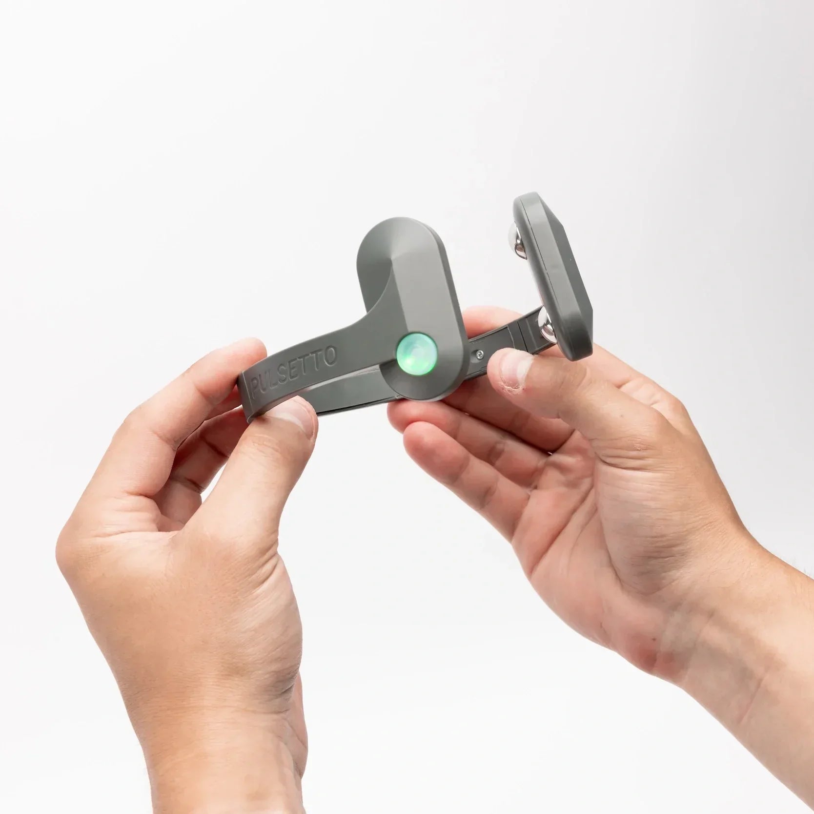
was followed by 1 min without stimulation. cVNS was delivered bilaterally using the Pulsetto
device (25 Hz, 100 μs, < 40 mA). atVNS and acVNS (10 Hz, 300 μs, 2-3 mA). Heart rate (ECG),
blood pressure (finger plethysmography), and a single-channel EEG (FP1 location) were
recorded continuously. Statistics included one-way ANOVA for repeated measures with post-hoc
t-tests for each experimental group. P < 0.05 was considered significant, 0.05 < P < 0.10 was
considered a trend.
Results:
Systolic blood pressure only decreased during cVNS (133.6±5.0 mmHg at baseline vs.
124.2±4.8 mmHg, n=7, P<0.05) but not in the time control or both auricular VNS groups. No
significant changes in heart rate were observed in any group. The amplitude of alpha waves in
the EEG increased only in the cVNS group (0.654±0.070 arb. units at baseline vs.
0.827±0.076 arb. units, n=7, P=0.06) but not in the time control or both auricular VNS groups.
No significant changes were observed for any other EEG waves.
Conclusion:
Non-invasive transcutaneous VNS reduced systolic blood pressure and increased
alpha waves in the EEG of the frontal lobe when the cervical vagus nerve was targeted
bilaterally, but not when the auricular branch of the vagus nerve was stimulated unilaterally.
Increased alpha waves are consistent with a more relaxed mental state which may have caused
the reduction in systolic blood pressure with cVNS potentially through decreased stress
hormone release.
Authors:
Rodela Ahmed, Andrea Coello, Aamani S. Pillutla, Gurpret E.E. Telwar, and Harald M. Stauss Department of Biomedical Sciences, Burrell College of Osteopathic Medicine, Las Cruces, NM
Running Head
Transcutaneous Cervical vs. Auricular VNS
For Correspondence
Harald M. Stauss, MD, PhD
Burrell College of Osteopathic Medicine
Department of Biomedical Sciences
3501 Arrowhead Drive
Las Cruces, NM 88001
Phone: 575-674-2327
E-mail: [email protected]
Table of contents:
Read more:
The use of non-invasive vagus nerve stimulation (Pulsetto™) as a post-exercise recovery strategy in male football players
Read StudyEvaluating Non-Invasive Vagus Nerve Stimulation for Pain Management in Bechterew Disease: A Pilot Study
Read StudyHave questions?
We’re here to answer! Contact us at: [email protected]